Course Content
MASTERING OCULAR ANATOMY: ADVANCED
Deepen your understanding of ocular anatomy as you delve further into the complex structures and functions of the eye. This lesson covers the eye's main parts and regions while developing your understanding of the principles of refraction and accommodation. You will also learn about the six extraocular muscles and gain valuable insight into how the eye moves.
0/6
MASTERING OCULAR ANATOMY: ADVANCED
About Lesson

Anatomy of the Retina

Chapter 3 of 5

The retina is the sensory body connected by the optic nerve to the brain. It contains 10 distinct layers of nerve cells, nerve fibers, light receptor cells and supporting tissue. Light receptor cells are either rods or cones. They send information to the brain via the nerve fibers and optic nerve.

Histology of the Retina

The retina is composed of 10 histologically different layers. Each layer contains a specific type of cells, neurons, structural cells or photosensitive cells. Click on the hotspots below to discover all the layers’ names. The top layers are the most anterior (toward the front of the eye), the bottom layers are the most posterior (toward the back of the eye).

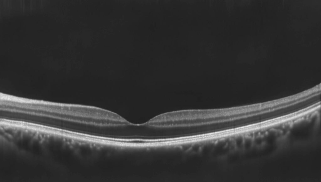
Retinal Layers OCT Image

We look at those structure clinically during an exam using an OCT. The image shows in vivo the 10 layers of the retina.

Pyramidal Architecture of Retinal Cells

Retinal cells are organized in a pyramidal structure: the information gets concentrated from the photoreceptors to the ganglion cells. For instance, 10 cones may connect to five bipolar cells, which connect to 1 ganglion cell. There is less concentration of information in the foveal region to preserve accuracy of information.

There Are 2 Types of Photoreceptors in the Human Retina: Rods and Cones

A radial section of a portion of the retina reveals that the ganglion cells (the retina’s output neurons) lie innermost in the retina, closest to the crystalline lens and the front of the eye. The photoreceptors (the rods and the cones) lie outermost in the retina against the pigment epithelium and choroid. Therefore, light must travel through the thickness of the retina before striking and activating the rods and the cones.

Rods Cones
130 million rods
7 million cones
Responsible for vision at low light levels (scotopic vision)
Active at higher light levels (photopic vision)
Do not mediate color vision, and have a low spatial acuity.
Capable of color vision and are responsible for high spatial acuity.

Optic Disc

The optic nerve enters the eyes at the posterior region. The contour of the optic nerve is known as the optic disc, where all retinal nerve fibers layers converge to form a bundle. It is important to note that the optic nerve does not contain photoreceptors, so any light focusing on that part of the retina cannot be seen. This results in a blind spot in a small area in the patient’s visual field where objects cannot be seen (scotomal. We are typically unaware layers converge to form a bundle. It is important to note that the optic nerve does not contain photoreceptors, so any light focusing on that part of the retina cannot be seen. This results in a blind spot in a small area in the patient’s visual field where objects cannot be seen (scotoma). We are typically unaware of those blind spots because they are small, and when both eyes are opened, they do not overlap.

Fovea

The fovea is the center of the macula. When someone looks at an object, they align the eye so the image falls on the fovea for best vision. The fovea has an anatomical pit due to the fact that not all of the ten retinal they align the eye so the image falls on the fovea for best vision. The fovea has an anatomical pit due to the fact that not all of the ten retinal layers are present in the fovea. That pit, or depression, is obvious during fundus examination. Light “concentrates” in the pit, leading to what is known as the foveal reflex: a small, bright spot of light on the fovea when light is shined inside the eye

Internal Limiting Membrane

Ganglion Cell Layer

Inner Nuclear Layer

Outer Nuclear Layer

Photoreceptor Layer

Retinal Pigment Epithelium

Choroid

Nerve Fiber Layer

Inner Plexiform Layer

Outer Plexiform Layer

External Limiting Membrane

Photoreceptor Outer Segment

Bruch's Membrane

Vitreal Side

Scleral Side

Retinal Pigment Epithelium

Rod Cells

Ganglion Cells

Cone Cells

Bipolar Cells

Retinal Nerve Fiber Layer